Watch More! Unlock the full videos with a FREE trial
Already have an account? Log In
Included In This Lesson
Study Tools
Access More! View the full outline and transcript with a FREE trial
Already have an account? Log In
Associated Lessons
Coronary Circulation is mentioned in these lessons
Transcript
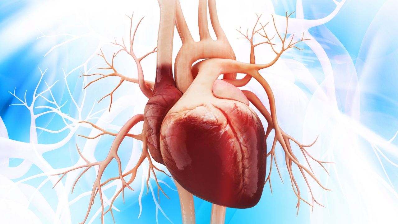
So, just as the heart is going to supply blood to the body, it also has to supply blood to itself. The heart is an enormous muscle and it requires a tremendous amount of cardiac output in order to just carry out its normal functions. So, because of that, the heart actually has its own system of arteries.
As you can see, the coronary arteries actually branch off of the aorta right above the aortic valve. That’s important to note, we’ll look at that in a second. On both sides of the heart you have a main coronary artery branching off - the Right Coronary Artery or RCA, and the Left Main Coronary Artery or LMCA. From there they each have smaller branches to supply the front and back of the heart. On the right we have the Right Posterior Descending which wraps around the back and the Right Marginal Artery which wraps around the front. On the left, we have the Left Anterior Descending or LAD that supplies the front left side of the heart and the Left Circumflex, often written LCX, which wraps around the back left side of the heart. You’ll notice there are more branches on the left, that’s because it is larger than the right. The main ones that are susceptible to blockage are the RCA, LMCA, LAD, and LCX - you’ll learn more about what a blockage means in the Myocardial Infarction lesson.
Now, it’s important to note what each of these coronary systems supplies blood to. On the left, it supplies the Left Atrium, Left Ventricle, and part of the Septum. On the right it supplies the Right Atrium, Right Ventricle, and part of the Septum as well, but it ALSO supplies the SA and AV node. So you can imagine that if someone had a blockage on the right side of their coronary perfusion, they might be susceptible to arrhythmias.
One more thing that’s important to note about how and when the heart perfuses itself. Remember how I said that the coronary circulation branches right above the aortic valve? Well if you think about systole and diastole. The heart contracts in systole and the Aortic Valve opens - when it does, it will briefly block the coronary circulation. Then the heart relaxes in diastole, the aortic valve closes, and blood flows through the coronary arteries. There is also a pause after diastole to allow filling of the ventricles, so this gives even more time for blood to flow to the coronary arteries. So, the heart perfuses the body during systole, and perfuses itself during diastole.
Time for some critical thinking! If my patient’s heart rate is extremely fast, what happens to their time in diastole? It decreases! Instead of Lub, Dub……..Lub, Dub…..Lub, Dub….. it becomes LubDubLubDubLubDub with no pauses in between. So if my heart perfuses itself during diastole, what happens to coronary perfusion in severe or prolonged tachycardia? It decreases dramatically!
It’s like pressing a water fountain button over and over really fast and expecting to be able to get a full drink of water. It’s not possible. You have to hold it. Without that diastolic pause, the heart struggles to perfuse itself.
In the ICU when we see a patient in a significant tachycardia (for example Supraventricular Tachycardia in the 160’s), who is stable so far, we all tend to say something like “can’t stay like that forever” or “his heart will give out eventually” because we know that the patient’s coronary perfusion is decreased significantly. So that’s just something I want you to know so that if you have a patient who’s been severely tachycardic for a long time, you will know how important it is to address the situation!
So remember, the coronary circulation is how the heart gets blood flow to itself. Both sides of the heart have a main coronary artery and then branches to cover the whole muscle. The heart perfuses the body during systole, but perfuses itself during diastole. And prolonged tachycardia can lead to decreased coronary perfusion and even a myocardial infarction.
I hope you learned something today! Go out and be your best self and as always, happy nursing!
View the FULL Transcript
When you start a FREE trial you gain access to the full outline as well as:
- SIMCLEX (NCLEX Simulator)
- 6,500+ Practice NCLEX Questions
- 2,000+ HD Videos
- 300+ Nursing Cheatsheets
“Would suggest to all nursing students . . . Guaranteed to ease the stress!”


